

- 17 Sep, 2024
- 0 Comments
- 6 Mins Read
SICS Cataract Surgery Training 2025
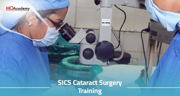
SICS Cataract Surgery Training
As compared to other techniques of cataract surgery and to attain competency in small incision
cataract surgery, there is a need for formal SICS Cataract Surgery Training which includes
wet lab and hands-on SICS step training under expert supervision.
An ophthalmologist well versed with the SICS technique can then perform the same effectively
and teach the technique to others who are interested in upgrading from the age-old technique
of couching to this technique and the modern generation aiming at avoiding blindness
from cataracts at an earlier stage.
SICS (Small Incision Cataract Surgery) is a microsurgical technique with minimal incision,
where the hard cataract is removed through an incision less than 3.2 mm and the IOL is implanted in the eye.
SICS cataract surgery is considered the technique of choice for cataracts in developing countries as it is fast,
cost-effective, and gives an outcome similar to that of phacoemulsification cataract surgery.
Overview of SICS Cataract Surgery Training
Small incision cataract surgery (SICS) has evolved as a sutureless, modified form of conventional
extracapsular cataract extraction surgery.
This Training has been designed keeping in mind the above need for a structured tutorial in learning SICS.
Starting right from the basic concepts, it aims to provide a complete overview of SICS cataract
surgery and intends to actively teach the steps in a simple and easily understandable manner.
Importance of Training in SICS Cataract Surgery
A lot of phaco surgeons tend to convert to phaco from SICS due to various intraoperative complications.
SICS surgery has a faster visual recovery rate when compared to phaco mainly because the energy
used in SICS is minimal and surgery is done by making use of internal wound constriction
and wound-assisted extraction of the nucleus.
Here again, in the early learning curve of SICS, many surgeons using the act of manual
small incision cataract surgery tend to do an extracapsular cataract surgery removing the nucleus out of the tunnel,
hence negating the same advantages of SICS to phaco.
The right training helps the surgeons to convert to SICS from ECCE and do SICS in the right way.
So the best way to get the desired results is through the right SICS Cataract Surgery Training.
SICS Cataract Surgery Training Curriculum
Surgeons will learn during this training:
1- Preoperative Preparation
Preparation of the patient must be carried out meticulously.
Trainees must learn the importance of a patient being still and steady throughout the surgery.
Sudden movement can cause the intraocular structures to shift,
making the surgery more difficult and increasing the chances of complications.
An anesthetized patient who is not in pain can be difficult to control,
and continuous reassurance is needed to prevent them from talking or moving.
It is quite useful to ask such patients to count aloud whenever they feel the urge to move; often the counting
itself provides adequate distraction.
2- Patient Assessment and Selection
A preoperative assessment can help in achieving the best possible visual outcome for the patient.
It helps to identify patients’ realistic visual needs and any special problems or goals
that need addressing during surgery.
It may also identify those patients for whom a referral to another professional or non-cataract
surgery would be more appropriate.
Patients with significant visual loss from cataracts and with little improvement in vision on pinhole
acuity are usually very happy about the results of their surgery.
The surgeon must be aware of patient expectations and should be able to predict visual potential
based on preoperative findings, to guide patient expectations towards realistic goals.
A full ocular examination should be performed, including a dilated funduscopy.
Thanks to modern cataract surgery techniques, patients can now be offered surgery
despite having coexisting ocular pathologies.
It is important, however, that the patient is fully informed about the increased risks
and the probable visual outcome following surgery.
Cataract surgery is generally contraindicated in cases of active ocular inflammation,
recent significant eye trauma, uncontrolled glaucoma, macular degeneration,
and when there are multiple sight-threatening ocular pathologies in the same eye.
3- Preparing the Operating Room
The doctor at the SICS Cataract Surgery Training needs to know the details of the operating room:
Step one: Ensure that the patient is in a comfortable position lying on the operating table.
– A near visual acuity chart may be useful to test patient vision.
Although unnecessary for routine cataract surgery cases, this can be useful for patients
with poor vision from a cataract combined with other ocular comorbidities.
– An operating microscope with a high-quality light source is necessary to visualize
the anterior segment and pupil clearly throughout the surgery.
– An air infusion system and a machine to remove fluid from the eye are essential.
Although it is possible to do SICS with a manual system for controlling intraocular pressure,
the ability to accurately set the infusion pressure and to finely control the fluid maintained
in the eye is crucial and is best done with an air infusion and bottle set.
– A machine to measure intraocular pressure should be available to test the IOP during
surgery and at the end of the case.
4- Surgical Techniques
Although there is a great deal of mix and match between various techniques of phacoemulsification and SICS,
we shall consider the basic technique of SICS as a hard nucleus extraction technique
without the use of a phaco machine.
– Incision Creation
This technique requires more surgical skill and at present has a longer learning curve than the scleral tunnel technique,
though the surgeon should not be dissuaded to learn the clear corneal incision method
as it is now considered the gold standard.
– Capsulorrhexis
A continuous curvilinear capsulorhexis is an essential step in extracapsular and intracapsular
cataract surgery as it facilitates adequate nucleus management in the former and aspiration
of residual cortical material in the latter.
– Nucleus Removal
– Intraocular Lens Implantation
An intraocular lens (IOL) is a lens implanted in the eye used to treat cataracts or myopia.
The most commonly used IOLs are fixed monofocal lenses that have one set focal point,
and multifocal lenses that work in a similar way to multifocal spectacles, providing focus at multiple distances.
In certain cases, where the natural lens or an IOL has been removed during surgery
(e.g. for infection with a perforating injury), it is possible to implant an IOL in the absence of a lens capsule.
Before the implantation of the IOL, it is important to thoroughly clean the viscoelastic from both the anterior
and posterior capsules to reduce the possibility of postoperative raised IOP and inflammation.
This can be achieved by aspirating the viscoelastic with the irrigation/aspiration probe or by inflating
and deflating the capsular bag with a cohesive viscoelastic to allow the viscoelastic
to be washed out of the capsular fornices.
5- Postoperative Care
An ideal setting for cataract postoperative care gives patients a comprehensive understanding of self-care,
dispels anxiety about the recovery, provides a point of contact during the recovery period,
and when necessary, facilitates rapid access to further care (complications).
During The SICS Cataract Surgery Training, Postoperative care is an essential component of cataract surgery.
The quality of postoperative care can be reflected in the incidence of postoperative complications.
Many factors can contribute to the delivery of optimum postoperative care,
including the strength of communication and the quality of educational input
at both the patient and the clinical levels.
Medication and Follow-up
During The SICS Cataract Surgery Training stress has been laid on the options available
for the management of complications.
Then, a more detailed discussion is initiated regarding the probable cause of the complication,
the different techniques available for its management, and the risk benefits involved with each technique.
If the surgical outcome is less than optimal, an early postoperative visit provides the surgeon
with an opportunity to detect the cause of sub-optimal visual recovery and correct
it before irreversible chronic changes occur.
Resolution of common clinical problems that the surgeon may encounter during the early postoperative phase,
such as corneal edema, moderate inflammation, and transiently raised IOP,
will usually result in a dramatic improvement in visual acuity.